Haemorrhoids, anal fissures or anal crypts and perianal fistulas? Whatever the diagnosis, it is important to remember that untreated lesions have an increased risk of malignancy.
Anoperative pathology
Who are we talking to?
To all those experiencing conditions such as haemorrhoids, anal fissures, anal crypts and perianal fistulas, haemorrhoidal thrombosis and thrombophlebitis or anoperian condylomatosis.
Why choose SKINMED experts®?
SKINMED® experts are at your side to find the most appropriate solution for the personalised treatment of these injuries/conditions. They offer personalised treatment plans, with state-of-the-art technology in the clinic that is extremely helpful in both dignostic accuracy and treatment.
Benefits of anoperative injury treatments in SKINMED®
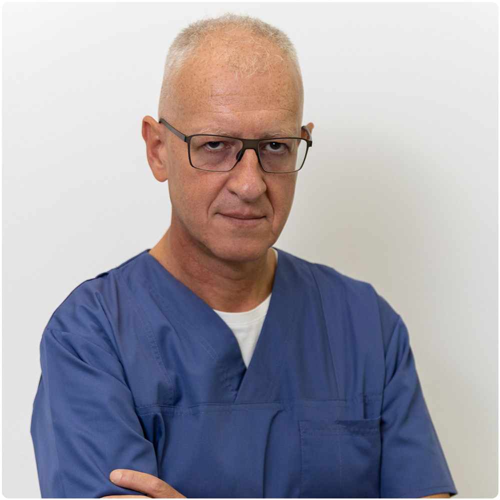
Team of experts with extensive experience and coordinated by Dr. Amalia Anghel
Doctors experienced in the latest news, treatments and technologies, with national and international interactions
State-of-the-art technologies
Modern and effective treatments and procedures
Proctology consultation
SKINMED® addresses the following conditions:
Diagnostic stage is extremely important because it determines both the condition and the treatment modality (conservative, interventional outpatient or inpatient). It is based on proctological consultation comprising:
Unlike colonoscopy, the proctological consultation does not require any special preparation, but it would be desirable for the accuracy of the examination that the patient has had a bowel movement prior to presenting for examination.
Although a diagnosis is generally established at the end of the examination, further investigations (blood tests, abdominal ultrasound, colonoscopy, possibly MRI) may be requested.
What types of anoperative conditions can we diagnose and treat in SKINMED®?
- Hemorrhoids
- Anal fissures
- Anal fissures and perianal fistulas
- Thrombophlebitis and haemorrhoidal thrombosis
- Anoperian condylomatosis
What are they and how do they manifest themselves?
Haemorrhoids are varicose dilatations of the veins in the internal and/or external haemorrhoidal plexuses (internal and/or external haemorrhoids, microscopic haemorrhoids). The veins in these plexuses are naturally more dilated and play an important role in the containment of faeces and gases. So, to some extent, every person has haemorrhoids and it is normal to have them. And not every hemorrhoid you encounter necessarily needs to be treated. But at some point, for various causes (functional/occupational, infectious, dysmetabolic, dysendocrine), the disease can occur.
The elements that determine the therapeutic indication are the bleeding (those of haemorrhoidal cause are, in general, the prerogative of internal haemorrhoids and are more or less light red in colour, separated from faecal matter and not mixed with it, in variable quantity, from a fine trace on toilet paper to a large quantity filling the toilet bowl), size (especially of external haemorrhoids), behaviour (periodic inflammatory phenomena, sometimes of high intensity, with variable response to conservative treatment, or intermittent anal prolapse, post defecation or after exertion).
Depending on size and behaviour (spontaneous reducible, digital or irreducible prolapse), internal haemorrhoids go through several evolutionary stages (grades) of the disease, numbered from 1 (least severe, where haemorrhoids are only slightly larger than "normal'') to 4 (irreducibly prolapsed internal haemorrhoids, not even manually).
How do we treat haemorrhoids?
As far as treatment in SKINMED® is concerned, it can be conservative or interventional. The greatest outpatient applicability is for rubber-band ligation of internal haemorrhoids. The procedure consists of placing a natural rubber band at the base of the mucosa of the haemorrhoidal mass. Necrosis of the apical haemorrhoidal tissue occurs within a few hours, partially including the vessels irrigating the haemorrhoid, leading to the removal of the haemorrhoidal mass within 2-7 days and then to the induction of fibrosis which will achieve healing (usually within 7 days). Internal haemorrhoids have no (or very little) tenderness, which means that they can be ligated in the usual way without causing pain. Elastic band ligation has eliminated the need for about 95 % of hemorrhoid surgeries. After ligation, dietary and exercise restrictions are minimal, but must be observed to ensure the success of the procedure and avoid possible complications.
When the problem is external haemorrhoids or haemorrhoidal marisci, elastic ligation cannot be applied. As for the interventional treatment of external haemorrhoids, given their sensitivity, a degree of anaesthesia is required. Laser therapy or radiofrequency approaches can be tried but excision of the external haemorrhoid is usually required.
What are they and how do they manifest themselves?
Anal fissure is an ulceration, initially linear, later oval, more or less superficial, causing local pain and anal sphincter hypertonia. It occurs in the distal portion of the anal canal, more commonly in the posterior and anterior commissures (9th and 3rd hours left lateral decubitus DLS), and can range from involvement of only the superficial skin layer to complete effacement of all integumentary layers.
Clinical manifestations are pain during and/or after defecation, sphincter contracture, bleeding (usually minor). Symptomatology may change as the anal fissure becomes chronic.
How do we treat anal fissures?
Initial treatment is conservative, aiming to break the vicious circle of "pain - sphincter hypertonia - pain". Nitroglycerin or Nifedipine ointments may be used, with or without topical corticosteroid, possibly with a local anaesthetic. Sitz baths in lukewarm (not hot or warm!) water after defecation usually help relieve symptoms. Medication with a relaxing effect on the smooth muscles (NoSpa) can also be administered. Classic anti-haemorrhoidal ointments are not effective in the treatment of anal fissures, and suppositories are contraindicated because they do not act on the fissure and their administration further traumatises an already injured area.
Botulinum toxin perilymph injection can be useful. A few days after injection, botulinum toxin produces a temporary, partial and reversible paralysis of the anal sphincter, which results in reduced to no pain. The effect of botulinum toxin lasts for 2-3 months, after which sphincter tone returns to normal. Since most patients present to the doctor at the time of chronic anal fissure, healing is almost impossible with botulinum toxin injection alone, so the 2-3 months should be used appropriately (continuation of topical treatment and interventional manoeuvres).
Interventional treatment is performed within SKINMED® and aims to clean the connective/cyst tissue at the level of the anal fissure with as little damage as possible to the sphincter apparatus. In general, these objectives can be performed under local anaesthesia, by cautery (with electrocautery or ultrasound). After the procedure, local treatment is continued, with monitoring, through regular check-ups to verify the level and correctness of healing. Given the possibility of changes in the anal area, it is sometimes necessary to apply additional therapy with own plasma (which is also performed within SKINMED®).
Sometimes, the hyperalgesic nature of the anal fissure (which does not even allow conservative treatment to be attempted) or the associated pathology, whether local (bulky haemorrhoids in the vicinity of the anal fissure, significant sclerotic retractions) or general (anticoagulant medication!), may mean that the only possible therapeutic approach is surgery.
What are they and how do they manifest themselves?
Perianal fistulas are, together with abscesses and phlegmons, part of anoperianal suppurative (infectious) pathology. In fact, the fistula is not a disease in itself, but a consequence of an abscess or phlegmon treated incorrectly or late. The origin of the abscess is in most cases in an anal crypt, a swallow's nest-like structure at the junction of the anal canal and the rectal ampulla, which is the opening of mucus-secreting glands. If these glands become infected (in the case of enterocolitis, or constipation, or the presence of foreign bodies), an abscess will form which will tend to progress to the tegument where it will rupture, discharge pus and form a fistula.
PERIANAL ABSCESS IS A MEDICAL AND SURGICAL EMERGENCY!
A perianal fistula is characterised by the presence of an opening(s) at the tegument through which pus is discharged. Sometimes the fistulous tract can be palpated. At other times, the fistula closes at the ends, abcesses, pain recurs, after which pus is discharged and the symptoms subside; the cycle repeats itself over time, with the number of external fistulous orifices multiplying and the tract branching. Sometimes (in the case of incomplete fistulas without external orifice), the purulent discharge is transanal. In addition to the existence of purulent secretions, the symptoms of fistulae may be associated with local pain, bleeding (anal, on defecation, or external, unrelated to defecation). The presence of internal haemorrhoidal pathology increases the risk of anal crypt infection.
How do we treat anal crypts and perianal fistulas?
To date, no conservative, non-interventional treatment has been identified that has a significant success rate.
As procedures that can be performed in SKINMED® under local anaesthesia, the following can be performed:
- fisttulectomy (total excision of the fistulous tract);
- fistulotomy (opening of the fistulous tract by its longitudinal sectioning followed by curettage of the tract and removal of scar tissue formed);
- fistulotomy with seton (an elastic band is fitted across the fistula, which gradually tightens and gradually performs the fistulotomy. It is the most time-consuming method, but the safest in terms of preserving sphincter integrity).
The choice of treatment method is made only after identifying the relationship of the fistulous tract to the sphincter apparatus (extra-, trans- or prasphincteric fistula). Sometimes this may require a local MRI examination.
In some cases, after fistula removal, it may also be necessary to perform a plasma therapy procedure (this procedure can also be performed at SKINMED®).
Haemorrhoidal thrombosis is the formation of a thrombus (blood clot) in a haemorrhoidal vein. If inflammation is associated with this, it is called haemorrhoidal thrombophlebitis. Unlike thromboses of veins in the limbs (especially the veins of the lower limbs), haemorrhoidal thromboses are not potentially life-threatening.
The clinical appearance is an external anoperianally located swelling (internal haemorrhoidal thrombosis is rare), usually with a purplish appearance, of variable size. The appearance of the integument and the level of pain are dependent on the intensity of the associated inflammatory phenomena. Since the symptoms are sometimes extremely similar to those of perianal abscesses (which, in unfavourable situations, can lead to life-threatening sepsis), any swelling of the anoperian region should be examined as soon as possible by the doctor to establish the correct diagnosis and the correct therapeutic attitude.
Haemorrhoidal thrombosis usually occurs as a result of an intense increase in pressure in the vein (straining to defecate - constipation or diarrhoea - , intense physical exertion, prolonged sitting, pregnancy or childbirth, especially natural childbirth) or local congestion (alcohol and/or spicy food abuse, sometimes perimenstrual). Haemorrhoidal disease is usually a contributory factor, especially in the case of recurrent thrombosis. Thrombophilic states are rare in these cases, but should be considered in the situation of repeated thrombosis without apparent cause.
How do we treat thrombophlebitis and haemorrhoidal thrombosis?
The mode of treatment depends on the clinical manifestations:
- small thromboses, with reduced painful symptoms, may only require conservative treatment (general venotonics and heparin ointments) until the clot is dissolved; if not, thrombectomy is recommended;
- thrombophlebitis is associated with venotonic and anti-inflammatory medication, classic antihaemorrhoidal ointments; after remission of inflammatory phenomena, conservative treatment can be continued or thrombectomy can be performed;
- in the case of large thromboses, with or without marked pain, or those with ulceration and bleeding or thromboses recurring in the same vein, the best therapeutic approach is thrombectomy performed under local anaesthesia, with removal of the thrombus and the vein in which it has developed, with recovery within about 2 days.
It should be noted that conservative treatment should be started in the first week after the onset of thrombosis, as its effectiveness becomes extremely reduced once thrombus fibrosis begins. Also, a vein reperfused by conservative treatment will have poorer quality walls than before the thrombosis, so recurrences may occur periodically; thrombectomy, by removing the affected vein, makes recurrence impossible.
Also called venereal vegetations, condylomatous lesions are caused by HPV (Human Papilloma Virus) infection and are characterised by the appearance of single or usually multiple wart-like lesions with a tendency to clump together. The lesions can appear on both the skin and mucous membranes. Transmission is usually sexual. In some cases the lesions are very similar to syphilitic lesions, hence the need for serological testing for syphilis. It is not obligatory for all people infected with HPV to have a wart.
A characteristic feature of condylomatosis is the tendency for rapid growth and extension to neighbouring regions. The appearance can become pseudotumoral, raising important treatment issues.
It should be noted that untreated lesions have an increased risk of malignancy, especially those with endocavitary location (anal canal, cervix, vagina).
In terms of anoperian location (condylomata may be found on the surface of the rectal mucosa/internal haemorrhoids, not just in the anal canal), there is no absolute association with a particular sexual orientation or practice.
If the patient is in a steady relationship, it is mandatory to check for HPV infection in the partner.
How do we treat anoperian condylomatosis?
Drug treatment may use ointments or solutions with trichloroacetic acid or podophyllin and is only applicable to external lesions, in relatively small numbers and sizes.
In the case of endoanal or perianal lesions of large number and size, the outpatient approach requires local anaesthesia and photothermocoagulation or electrocautery or radiofrequency excision may be used (in several treatment sessions). The interval between sessions depends on the speed of tegument recovery. In the case of lesions located on the internal haemorrhoids, it is preferable to remove the haemorrhoids with elastic ligatures.
Pseudotumoural lesions require in-hospital surgical treatment, sometimes with associated plastic surgery.
Regardless of the method of treatment, the patient will be followed up post-surgery by the SKINMED® doctor to minimise the risk of recurrence.
It is useful to combine immunomodulatory therapy (Isoprinosins), possibly with HPV vaccination.
Doctors who perform these treatments:
SKINMED experience and technology®,
for health and youth!
Advantages SKINMED®
Experience
- Over 19,000 patients treated in the clinic
- Over 300 new patients every month
- Over 20000 treatments per year
Credits
- FOTOFINDER-accredited Centre of Expertise in the Diagnosis and Treatment of Skin Cancer
- ALMA LASER accredited laser treatment centre of expertise
- ALLERGAN accredited centre of expertise in Aesthetics and Medical Injecology
Safety
- Only world-class accredited technologies

