Our skin is like a map. It reflects our state of health and we are each responsible for its integrity.
Dermato-oncological consultations
Total body scan
Who are we talking to?
Dermato-oncological consultations are necessary for people diagnosed with malignant melanoma or severe skin diseases, as well as for anyone who suspects a condition. They must be correctly diagnosed for effective treatment. During this consultation, a total body scan is performed using FOTOFINDER® technology, which provides a certain, early diagnosis of melanoma, an extremely aggressive type of cancer. Sometimes, unfortunately, patients arrive at the practice for specialist screening far too late. This is why we always emphasise that medicine also has a preventive role, fulfilled by early visits to the practice.
Why choose SKINMED® experts?
Because we are a FOTOFINDER®-accredited Expert Centre for the Diagnosis and Treatment of Skin Cancers, with the most effective and safe resources to keep your skin healthy and beautiful! Our experts specialize in the diagnosis and treatment of skin conditions, with the most effective diagnostic and treatment technologies available to achieve the best and safest results for the patient. We also strive to explain to our patients the importance of caring for their skin and provide them with the resources to do so. We are committed to helping patients feel comfortable during their visit to the clinic, and our treatments are performed in complete safety, following current medical guidelines.
Benefits of dermato-oncological consultations in SKINMED®
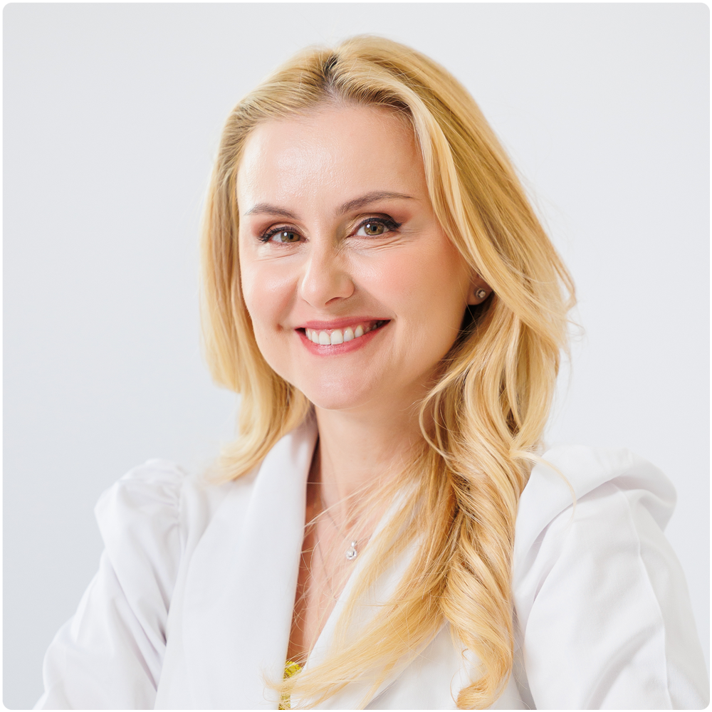
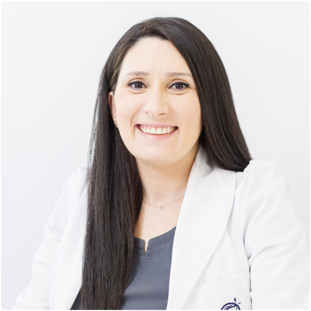
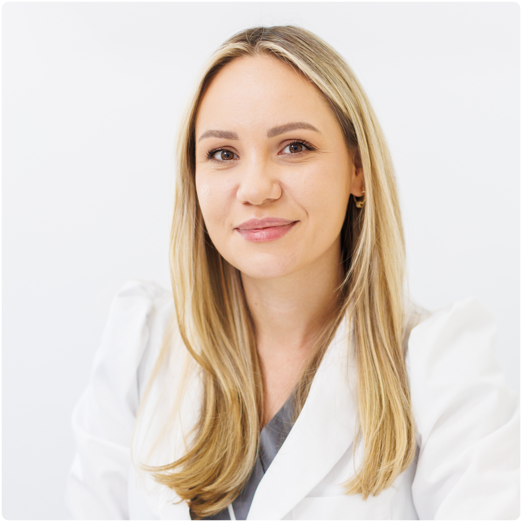
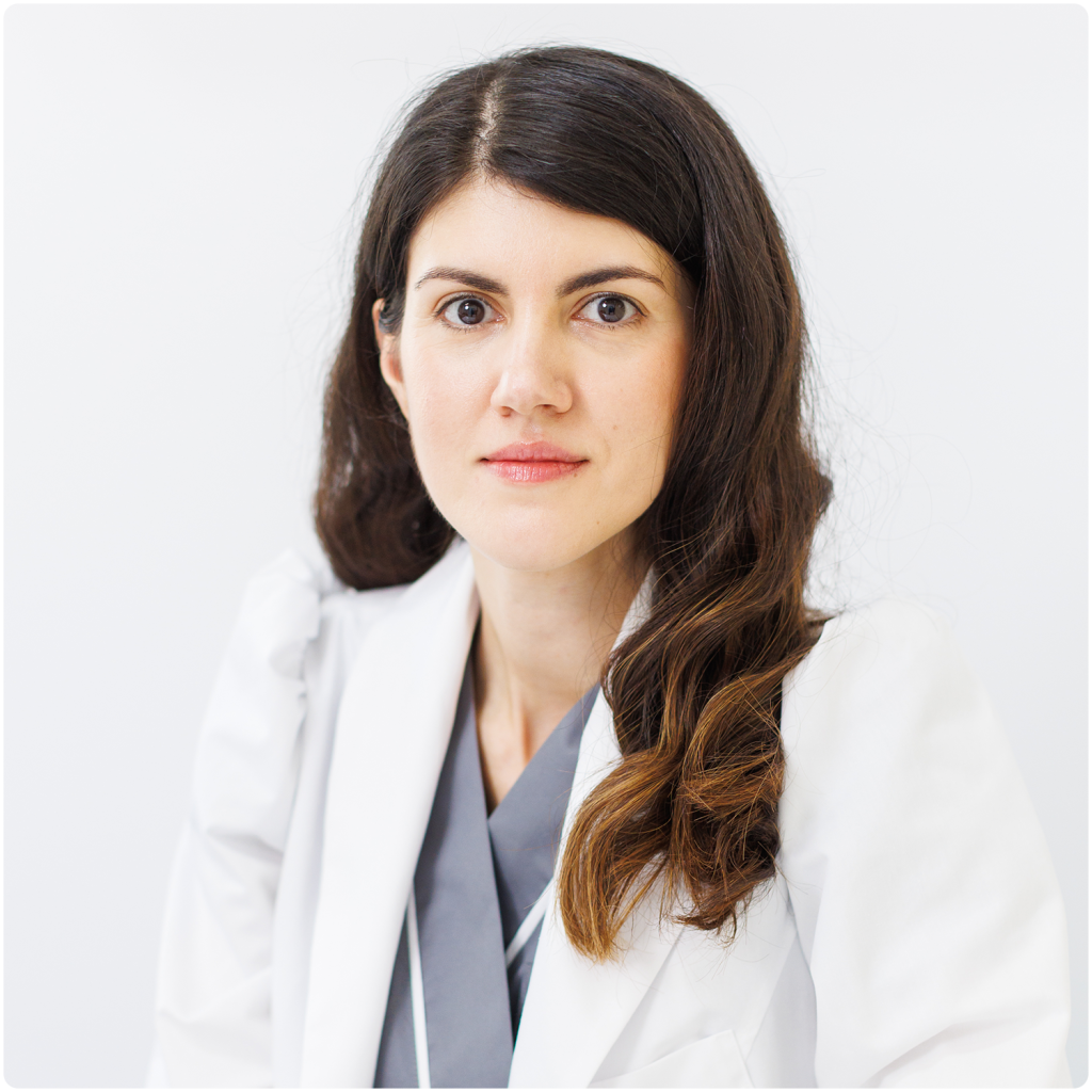
Team of professionals, coordinated by Dr. Amalia Anghel
Doctors trained in the latest treatments and technologies
Experts with national and international specialisations
State-of-the-art technologies
Modern and effective treatments and procedures
What is mole scanning with FOTOFINDER®?
Our nevus (mole) mapping system on the skin surface, developed exclusively by FOTOFINDER®, is designed to analyse in depth the structure of a nevus in order to detect any potential melanoma that cannot be seen on clinical examination. The image taken is basically a generalised dermatoscopy, and the image can be enlarged to view possible incipient malignant lesions in detail.
A reminder that melanoma, the skin cancer with the highest associated mortality, is difficult for the untrained eye to detect. At the same time, early diagnosis increases the cure rate.
SKINMED EXPERT Doctors®


SKINMED experience and technology®, for flawless looking skin!
Advantages SKINMED®
Experience
- Over 19,000 patients treated in the clinic
- Over 300 new patients every month
- Over 3,500 new patients annually
- Over 20,000 treatments per year
Credits
- FOTOFINDER-accredited Centre of Expertise in the Diagnosis and Treatment of Skin Cancer
- ALMA LASER accredited laser treatment centre of expertise
- ALLERGAN accredited centre of expertise in Aesthetics and Medical Injecology
Safety
- Only world-class accredited technologies
Other services you might be interested in
Frequently asked questions
What is melanoma?
Melanoma is a type of skin cancer that starts in the melanocyte, the cell that produces melanin, the pigment found in the skin, eyes and hair. Melanocytes are the cells that form the basis of moles. Although the majority of moles do not undergo malignant transformation, they represent the pre-existing lesion associated with melanoma, 1:5 cases, the remainder are new-onset lesions easily detectable by whole-body dermoscopy.
The good news is that detecting melanoma at early stages is associated with a cure rate of over 95%.
What is FOTOFINDER® scanning?
The FOTOFINDER® ATBM MASTER full skin surface scan (map) means the analysis of all lesions on the whole body. We study all the patient's skin lesions from head to toe, and in addition, our system monitors even the smallest changes and the evolution of the lesions over time.
Which patients should have their nevi mapped with FOTOFINDER®?
If you meet any of the conditions below, you associate an increased risk of developing melanoma skin cancer and should consider having a dermatoscopic map of the moles on your body:
- Multiple nevi (over 100 throughout the body)
- Several dysplastic/atypical nevi (more than 50 throughout the body)
- Family/personal history of melanoma
- Melanoma or other previously diagnosed skin cancer
- Previous diagnosis of neural dysplasia
- Large nevi (diameter over 5 cm) and/or atypical, asymmetrical shape
- Noticeable changes in pre-existing or newly developed nevi
- History of severe sunburn in childhood or adolescence
- History of artificial tanning (solar)
- Very light/light skin phototype (Fitzpatrick I / II)
How is the dermatoscopic nevus map performed?
At the simplest level, the dermatoscopic nevus map consists of a series of photographs showing the location and dermatoscopic appearance of the nevus. Nowadays, the safest way to do this is with a set of photographs showing the whole body surface so that we can visualise all the melanocytic skin lesions.

